The human foot is an incredible feat of biological engineering since it supports the whole human body. The foot is a sophisticated system made up of bones, joints, and muscles that allow us to stand, walk, run, and do other actions necessary for everyday living.
Discover the amazing complexity of the human foot as we count the bones, examine the joints, and learn about the muscles that all work together to keep us upright and mobile. Let’s read below “How Many Bones Joints and Muscles Does a Human Foot Have”:-
Bones of the Human Foot
The adult human foot consists of 26 bones, which are divided into three main regions: the hindfoot, midfoot, and forefoot.
Hindfoot:
-
- Calcaneus (Heel Bone): The largest bone in the foot, the calcaneus forms the foundation of the hindfoot and provides crucial support for body weight.
- Talus: This bone articulates with the tibia and fibula to form the ankle joint, allowing for up-and-down movement of the foot.
Midfoot:
-
- Navicular: A boat-shaped bone located in the middle of the foot, the navicular plays a key role in maintaining the foot’s arch.
- Cuboid: Situated on the outer side of the foot, the cuboid connects with the metatarsals and the calcaneus.
- Three Cuneiform Bones (Medial, Intermediate, Lateral): These small wedge-shaped bones articulate with the metatarsals and play a crucial role in supporting the arches of the foot.
Forefoot:
-
- Metatarsals (I-V): Five long bones extending from the midfoot to the toes, the metatarsals provide support and serve as the base for the toes.
- Phalanges (Proximal, Middle, Distal): The toes are comprised of 14 phalanges (toe bones), with each toe (except the big toe) having three phalanges.
Joints of the Human Foot
The foot joints can be categorized into three main types: synarthroses, amphiarthroses, and diarthroses.
Synarthroses (Immovable Joints):
-
- Interosseous Membrane Joints: Found between the tibia and fibula, these joints provide stability and minimal movement.
Amphiarthroses (Slightly Movable Joints):
Midfoot Joints: These joints, located between the cuneiform bones, cuboid, and navicular, permit limited movement, thereby enhancing the overall flexibility of the foot.
Diarthroses (Freely Movable Joints):
Ankle Joint: Formed by the articulation of the tibia, fibula, and talus, the ankle joint enables dorsiflexion (lifting the foot) and plantarflexion (pointing the foot).
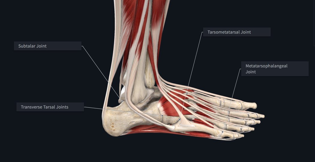
Subtalar Joint: Located between the talus and calcaneus, this joint facilitates inversion (turning the sole inward) and eversion (turning the sole outward) movements.
Midtarsal Joints: These joints allow for movement between the hindfoot and midfoot, contributing to the foot’s adaptability to different surfaces.
Metatarsophalangeal (MTP) Joints: Connecting the metatarsals to the proximal phalanges, these joints permit flexion and extension of the toes.
Interphalangeal Joints: Found between the phalanges, these joints enable bending and straightening of the toes.
Muscles of the Human Foot
The intricate movements of the foot are orchestrated by a network of muscles that work together to provide stability, support, and the ability to generate force. The muscles of the foot can be broadly categorized into intrinsic muscles, which are contained within the foot, and extrinsic muscles, which originate outside the foot.
Intrinsic Muscles:
Dorsal Interossei: Located between the metatarsal bones, these muscles abduct the toes (spread them apart).
Plantar Interossei: Situated between the metatarsal bones, these muscles adduct the toes (bring them together) and assist in flexion.
Lumbricals: Connecting the tendons of the flexor digitorum longus, these muscles flex the metatarsophalangeal joints and extend the interphalangeal joints.
Flexor Hallucis Brevis: Located on the plantar surface, this muscle contributes to flexion of the big toe.
Abductor Hallucis: Positioned on the medial side of the foot, this muscle abducts the big toe.
Flexor Digiti Minimi Brevis: Found on the lateral side, this muscle flexes the pinky toe.
Adductor Hallucis: Assisting in adduction of the big toe, this muscle is located on the plantar surface.
Extrinsic Muscles:
Gastrocnemius and Soleus: These muscles, collectively known as the calf muscles, play a crucial role in plantarflexion of the ankle.
Tibialis Anterior: Situated on the front of the leg, this muscle dorsiflexes the ankle and inverts the foot.
Peroneus Longus and Brevis: Located on the lateral side of the leg, these muscles evert the foot and assist in plantarflexion.
Flexor Digitorum Longus: Positioned along the posterior aspect of the leg, this muscle flexes the toes and plantarflexes the ankle.
Flexor Hallucis Longus: Running down the back of the leg, this muscle flexes the big toe and plantarflexes the ankle.
Extensor Digitorum Longus: Situated on the anterior side of the leg, this muscle extends the toes and dorsiflexes the ankle.
Extensor Hallucis Longus: Running along the front of the leg, this muscle extends the big toe and dorsiflexes the ankle.
FAQs
How Many Bones Joints and Muscles Does a Human Foot Have?
Complex Foot Structure:
- Comprises 33 joints, 26 bones, and over a hundred muscles, tendons, and ligaments.
Collaborative Functionality:
- Works collectively to support weight, facilitate movement, and transmit force efficiently.
How many muscles are in your foot?
Muscular Composition of the Human Foot:
- Total of 29 muscles are linked to the human foot.
- 19 muscles are intrinsic to the foot itself.
Conclusion
The flexibility and adaptability afforded by this elaborate framework make it possible to engage in a broad variety of pursuits. It is important for non-medical workers to understand foot anatomy as well. I hope you like reading “How Many Bones Joints and Muscles Does a Human Foot Have?”